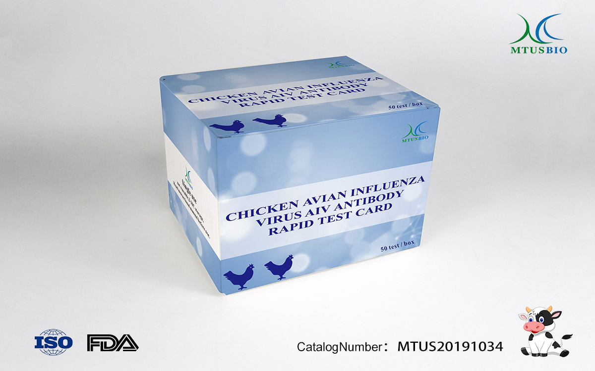
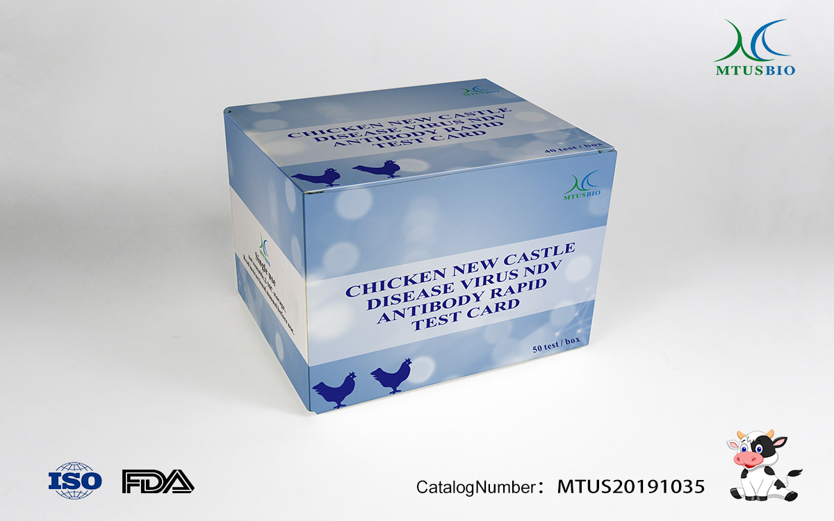
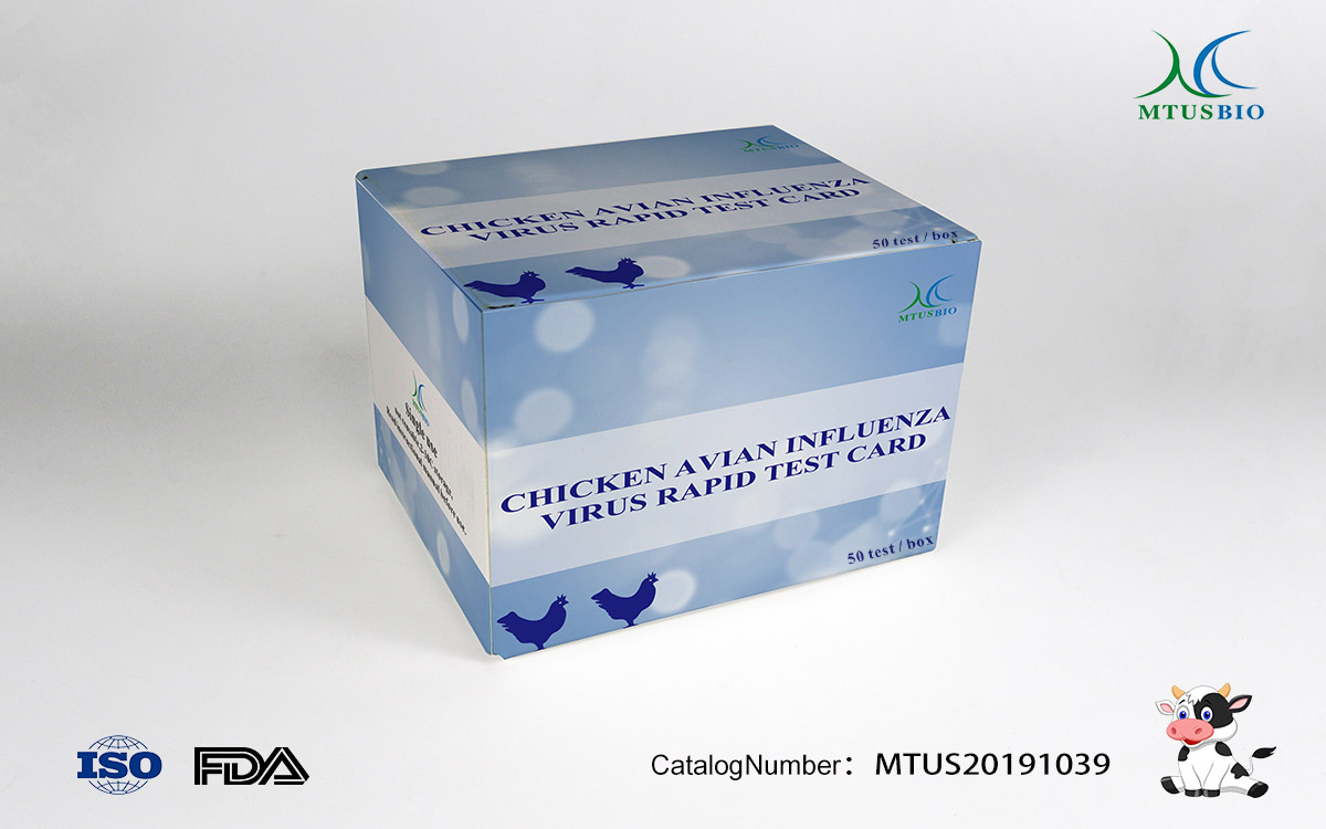
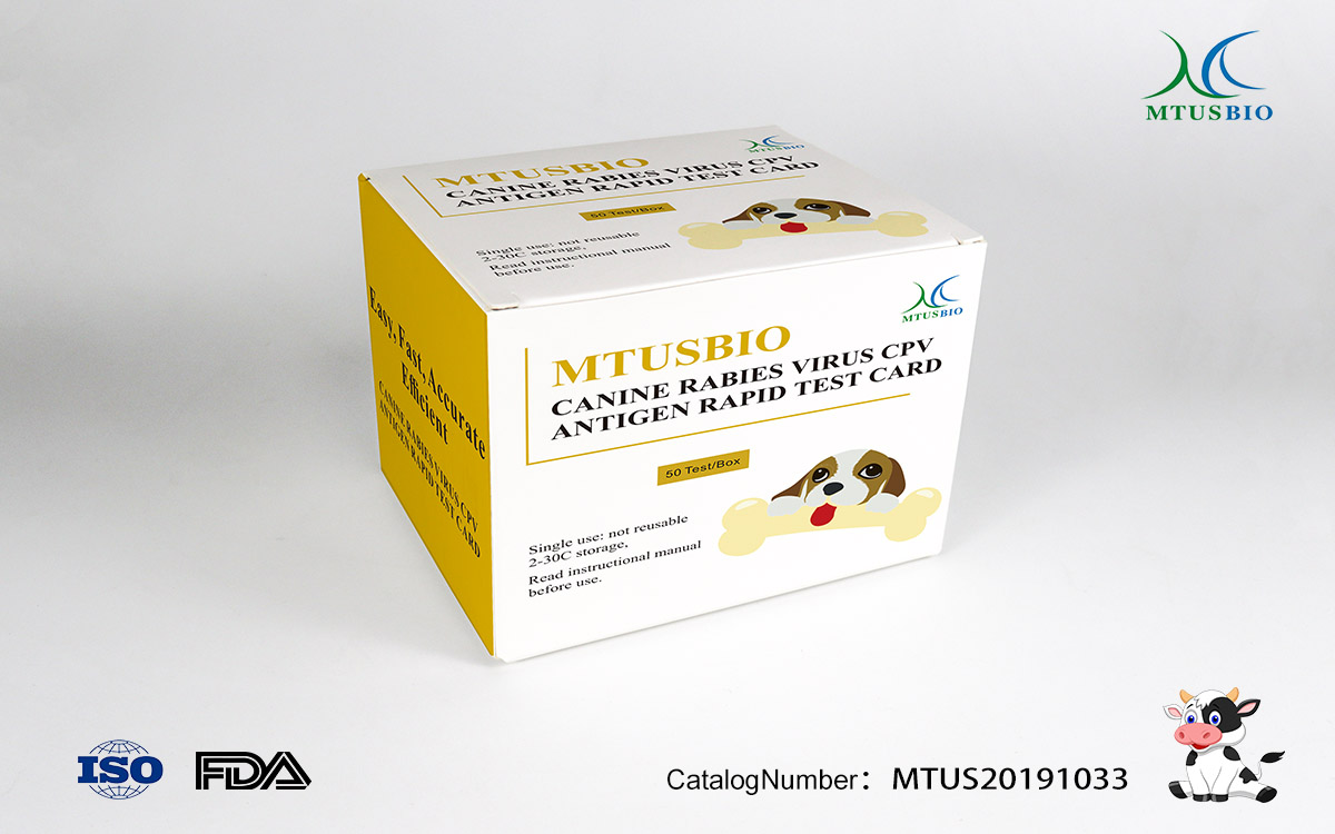
[Principle]
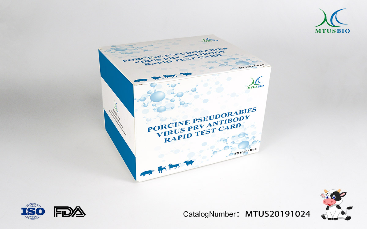
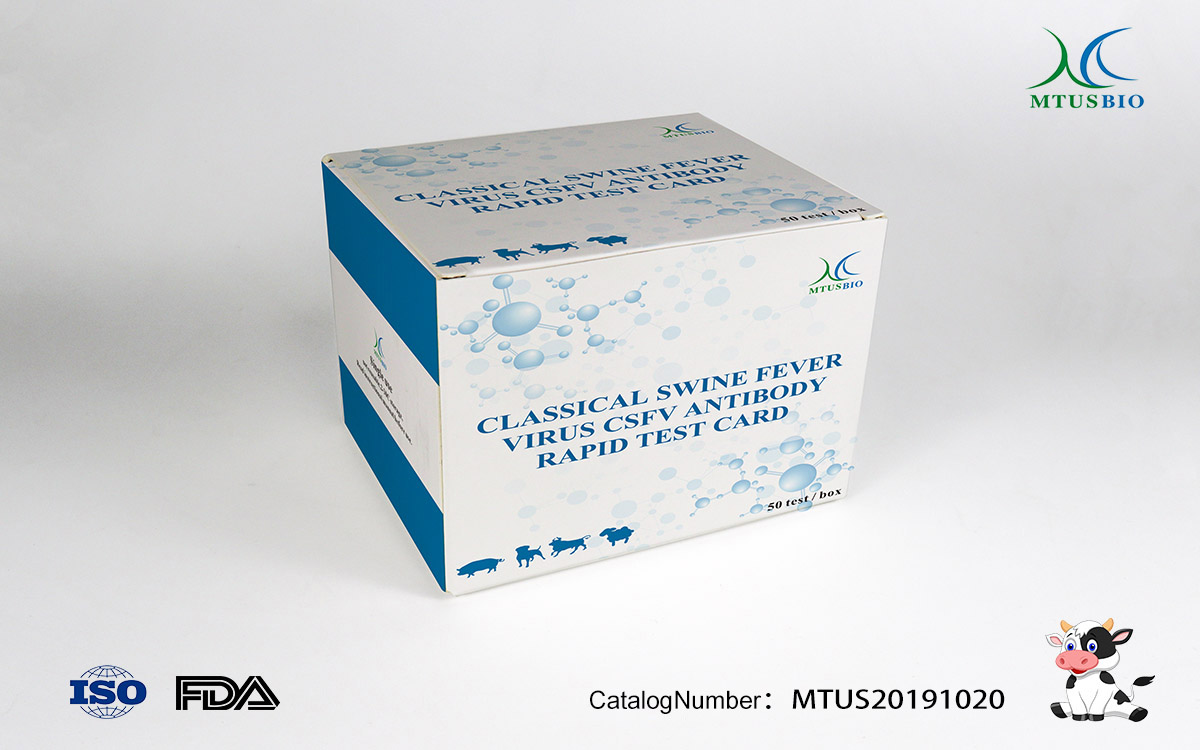
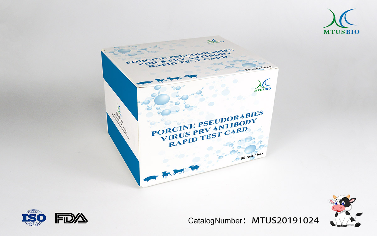
This reagent is made by the principle of colloidal gold immunochromatographic test (GICA). After the sample is added to the sample well, it moves along the chromatographic membrane together with the colloidal gold label. The antigen on the line binds and shows a magenta color. If no PRV antibody is present in the sample, no color response is produced.
[Kit composition]
No. | Name | 50T / box |
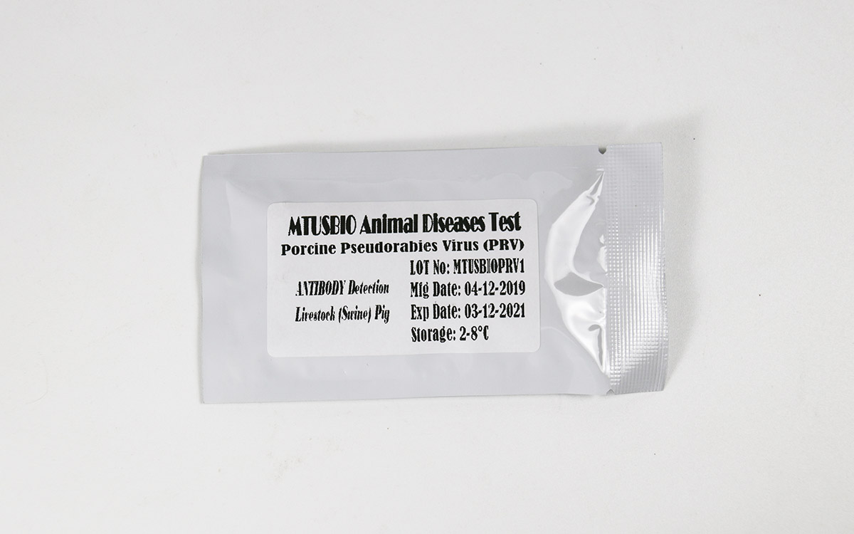
1. | test cards (including straws) | 50 |
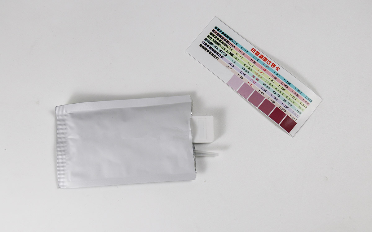
2. | antibody titer colorimetric card | 1 |
3. | instruction manual | 1 |
[Storage and expiry date] Store in a cool and dry place (2-30 ° C), valid for 24 months.
[Sample requirements]
1. This test card is only suitable for testing pig serum.
2. Serum: Take 2-3ml of blood according to the conventional method and place it in a clean and dry test tube. Let it stand for about 1 hour. After the blood is coagulated, centrifuge at 4000 rpm for 10 minutes. Serum is naturally precipitated), Eand the serum is separated. Require serum to be clear, no hemolysis, no pollution. Serum samples can be stored at 2-8 ° C in the short-term and at -20 ° C in the long-term.
【Experiment method】
1. Tear off the aluminum foil packaging bag of the test card, remove the test card, and place it on a flat, clean surface.
2. Pipette the prepared clear sample supernatant with a matching pipette, and slowly add 2-3 drops (about 60ul) into the sample well vertically and slowly.
3. Leave at room temperature for 10-20 minutes to judge the results. Results longer than 30 minutes are invalid.
1. Negative: In the observation hole, only a purple line appears in the control line area (C).
2. Positive: In the observation hole, a purple line appears in both the test line area (T) and the control line area (C). The higher the antibody titer, the darker the test line (T).
3. Invalid: In the observation hole, no purple line appears in the control line area (C).


